
Protein Structure & Nucleic Acids
Levels of protein structure; directionality of DNA and RNA.
The big idea: structure determines function
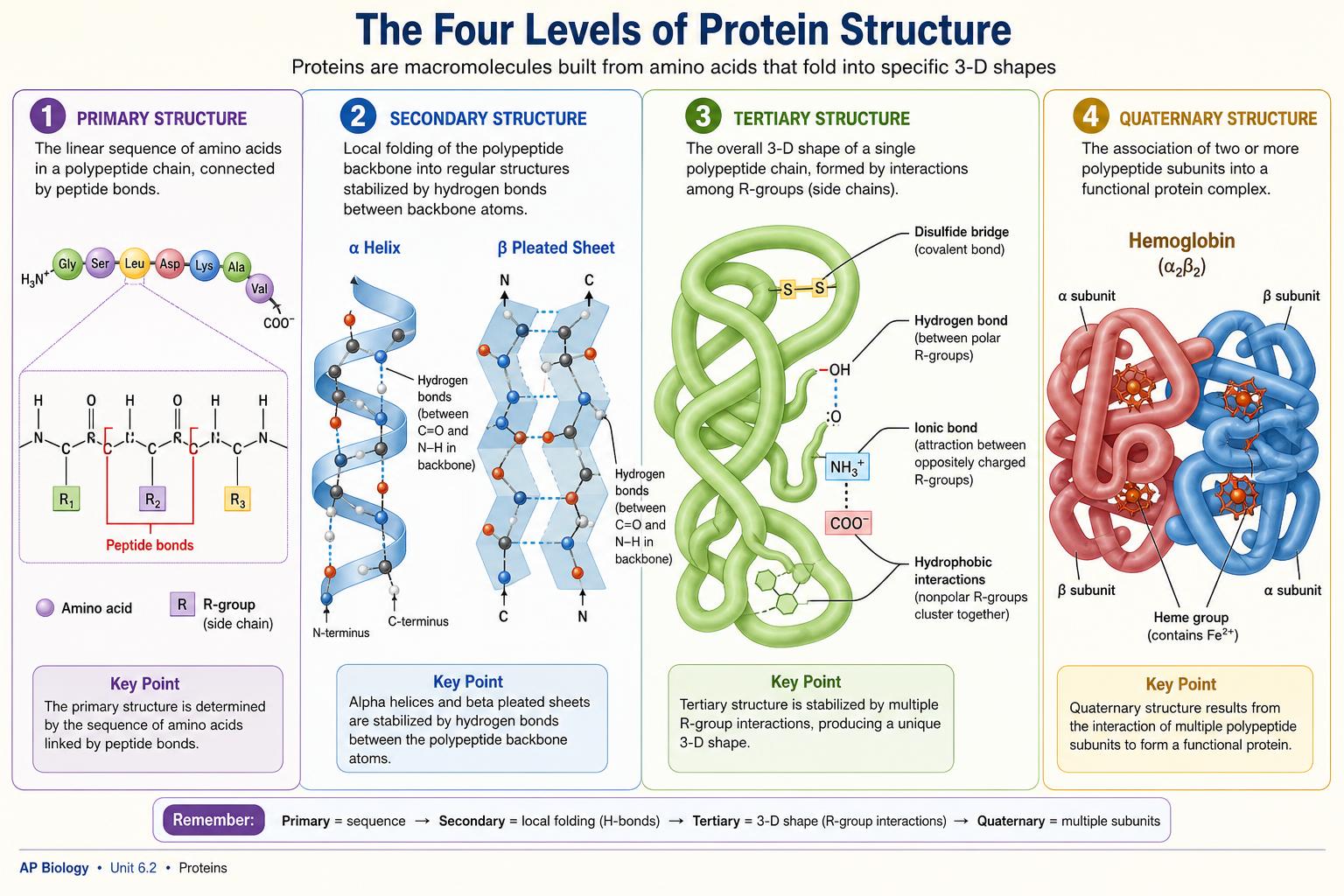
A protein's amino acid sequence strongly influences how it folds, and its 3D shape determines what it can do. Environmental conditions (pH, temperature, ionic strength), molecular chaperones, chemical modifications, and interactions with other molecules can also affect its final structure and function.
The classic example of sequence-driven change is sickle-cell hemoglobin: a single glutamic acid → valine substitution at position 6 of the β-chain replaces a polar residue with a nonpolar one. The new hydrophobic patch makes hemoglobin molecules clump under low O₂, deforming red blood cells.
Primary structure
The linear sequence of amino acids in a polypeptide, held together by peptide bonds. Read from N-terminus to C-terminus. The sequence is determined by the mRNA codon order, which traces back to DNA.
Secondary structure
Local folding patterns stabilized by hydrogen bonds between backbone atoms (N–H and C=O), NOT R-groups. The two patterns to know are α-helices (coiled, like a corkscrew) and β-pleated sheets, which consist of neighboring polypeptide segments held together by backbone hydrogen bonds. The strands in a β-sheet may be parallel or antiparallel.
Tertiary structure
The overall 3D fold of a single polypeptide, driven by interactions between R-groups: the hydrophobic effect (nonpolar R-groups clustering in the protein core away from water), hydrogen bonds, ionic interactions between charged R-groups, van der Waals interactions, and disulfide bonds (covalent S–S bonds between two cysteines).
In many soluble proteins, the hydrophobic effect is a major driver of folding because nonpolar R-groups tend to become buried away from water. Hydrogen bonds, ionic interactions, van der Waals interactions, and disulfide bonds can also help stabilize tertiary structure — the dominant force depends on the specific protein and its environment.
Quaternary structure
When two or more polypeptide chains assemble into a single functional unit. Hemoglobin is the canonical example: 4 subunits (2 α + 2 β), each with a heme group, working together to bind 4 O₂ molecules cooperatively.
Denaturation
Heat, extreme pH, or certain solvents disrupt the non-covalent interactions that hold tertiary and quaternary structures together. The polypeptide unfolds and loses function — the primary structure (peptide bonds) is usually still intact.
Frying an egg denatures its proteins irreversibly; this is also why high fevers are dangerous (enzymes start to lose shape) and why pepsin works at stomach pH but is inactivated in the small intestine.
Nucleic acid structure quick reference
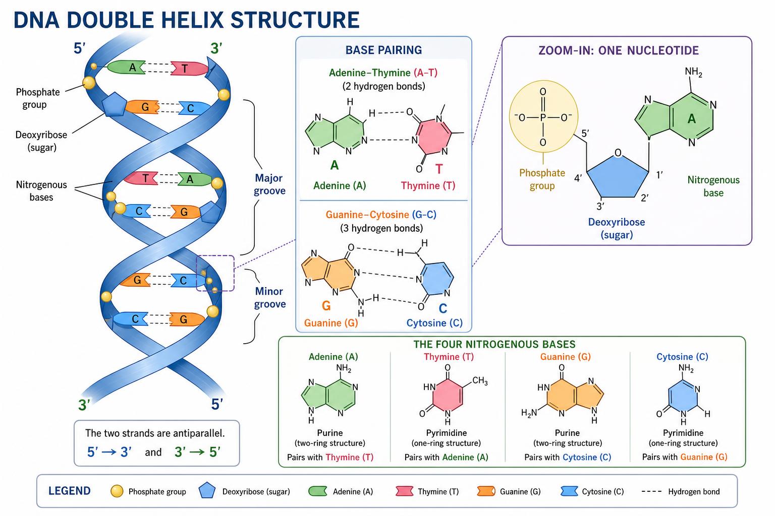
- DNA: deoxyribose sugar, double-stranded, bases A–T and G–C, antiparallel, runs 5'→3'.
- RNA: ribose sugar, usually single-stranded, U replaces T, several types (mRNA, tRNA, rRNA).
- Hydrogen bonds: 2 between A–T, 3 between G–C → G–C-rich regions are harder to separate.
- Phosphodiester bonds link the 3' OH of one nucleotide's sugar to the 5' phosphate of the next.
Key terms
Quick definitions to lock in before the exam.